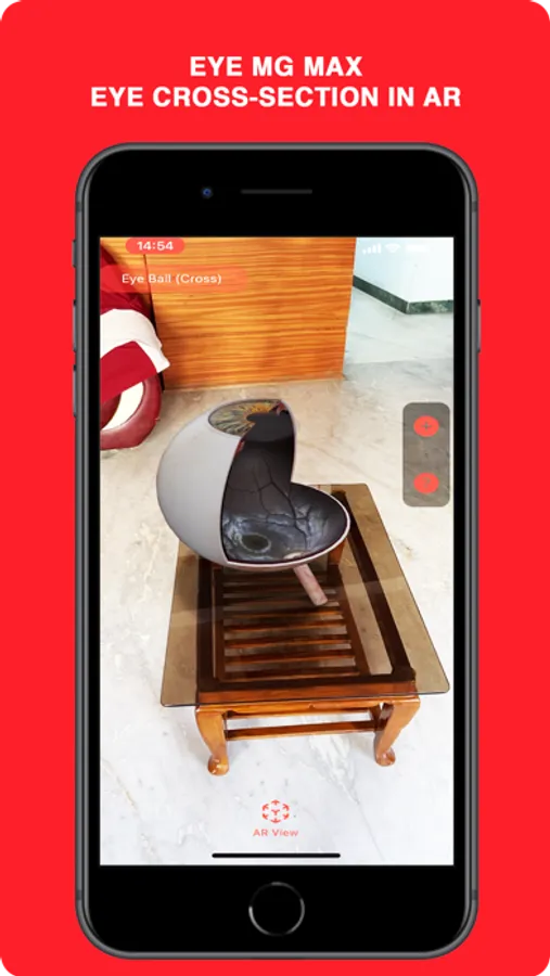
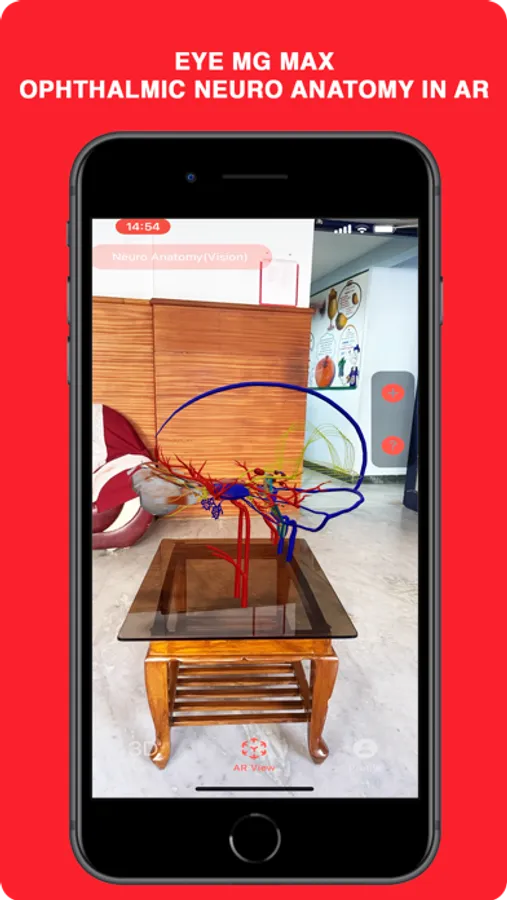
About Eye MG Max
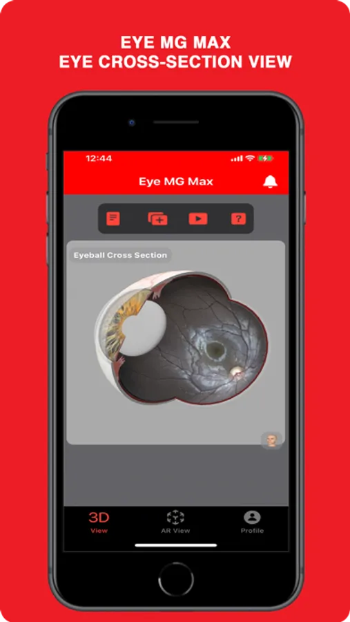
In Eye MG Max, a 3D eyeball model with TrueColor confocal fundus image, and all structures related to ophthalmology are provided in a user-friendly interface. In this application multiple views, along with transparency for viewing the structures passing through another model, free camera mode, annotated modes, customized zoomed views and videos related to any ophthalmic pathology is provided. Thus, providing a 3D atlas at the user’s fingertip for comprehensive learning.
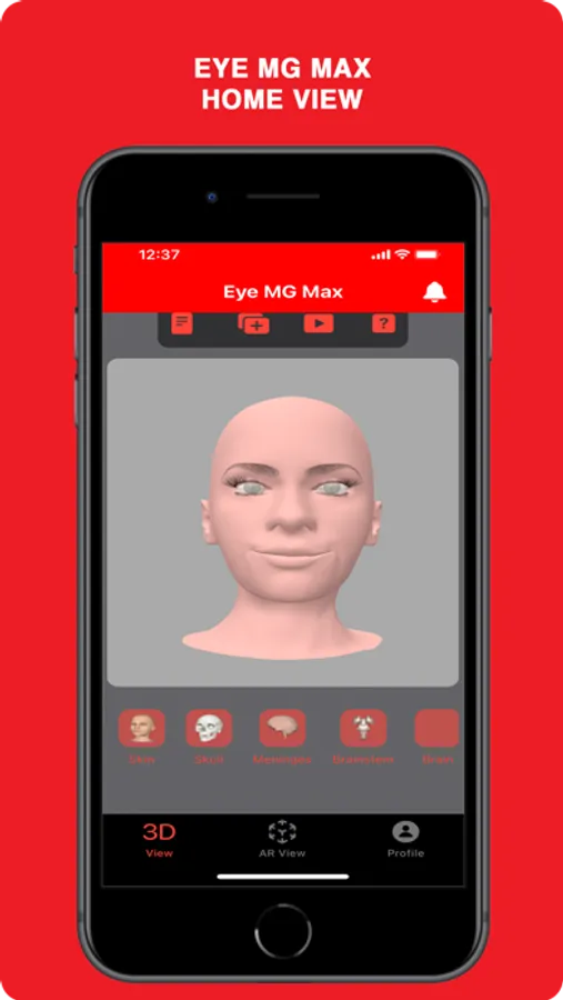
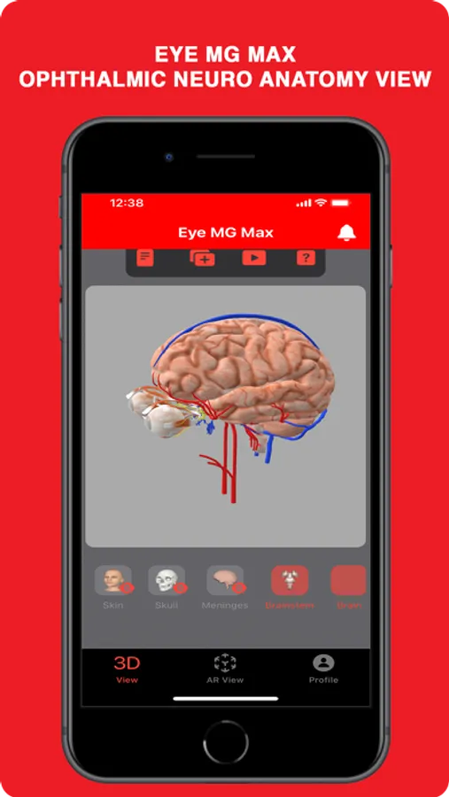
Free Mode: The user can explore the ‘3D View’ provided in the app, with Hide/Unhide option for various anatomical parts:
- Skin
- Skull
- Meninges
- Brainstem (BS)
- Brain
- Cranial Nerves (CNs)
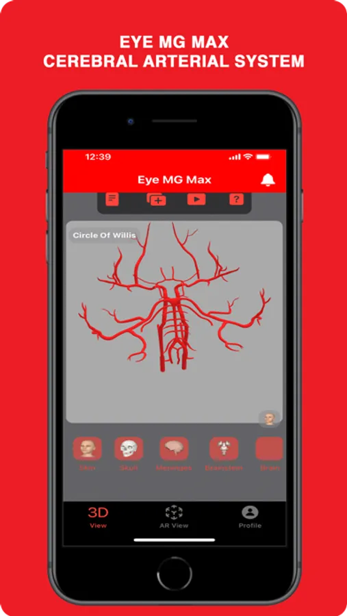
- Circle of Willis (CoW)
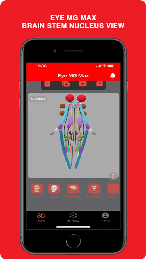
- Brainstem Nucleus (BS Nuclei)
- Optic Pathway (Optic Path)
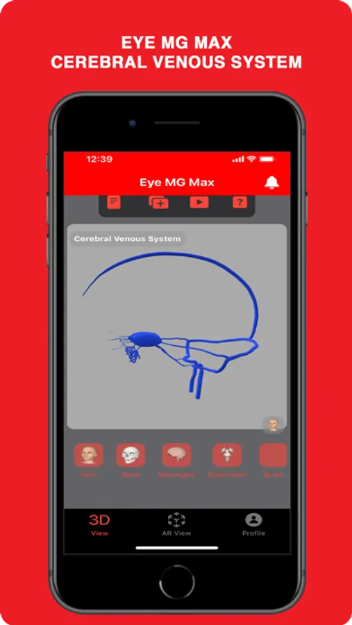
- Cerebral Venous System (CVS)
- Eyeball (Eye)
- Extraocular Muscles (EOM)
Users can explore these structures separately from the main menu:
- Eyeball
- Optic Pathway
- Brainstem Nucleus
- Cranial Nerves
- Cerebral Venous System
- Circle of Willis
- Skull - Supraorbital Fissure
AR:
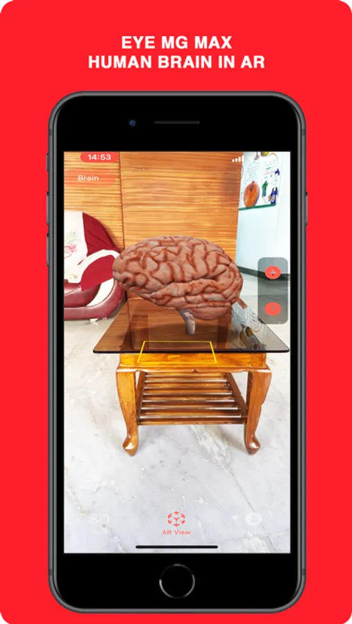
AR is a view of the real, physical world in which the elements are enhanced by a computer-generated overlay. AR requires a real world entity that can be overlayed on. For example, the face of a person, or a particular design or QR code. Sometimes even just a plane such as the ceiling, floor or wall would be enough to get the computer generated overlay (augmented world) onto the real world. In Eye MG Max, we just make use of a plane recognition system to overlay ophthalmic concepts and anatomical 3d models over the real world that would help ophthalmic residents and neophytes to experience the real-time structures.
Requirements for AR:
- iPhone 11 / iPad 2nd Gen or later
- iOS 13 / iPad OS or later
Permissions:
- Camera Permission for AR
Other Features:
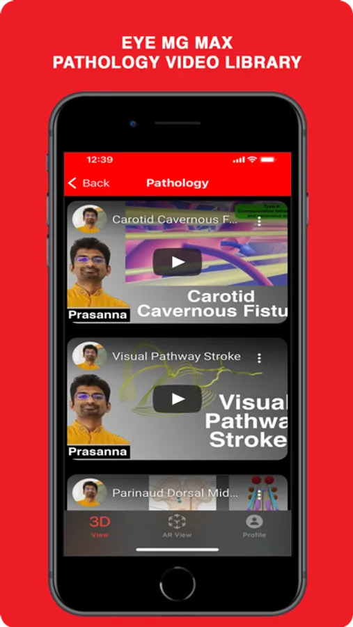
Video Gallery: Videos related to various pathologies are given here.
Help Section: Guide on how to use the ‘Free mode’ using Touch.
