About AGLV1199 CORTES ANATÔMICOS
1. OBJECTIVE
In this laboratory, you will study important aspects of diagnostic imaging, taking into account fundamental concepts of anatomy and imaging, as biomedical professionals working in this area must have extensive knowledge of these sciences in their professional practice. The human body is made up of cells, tissues and organs that are grouped into organic systems. Although the body presents similarities between individuals of the same species, we can say that each individual is unique.
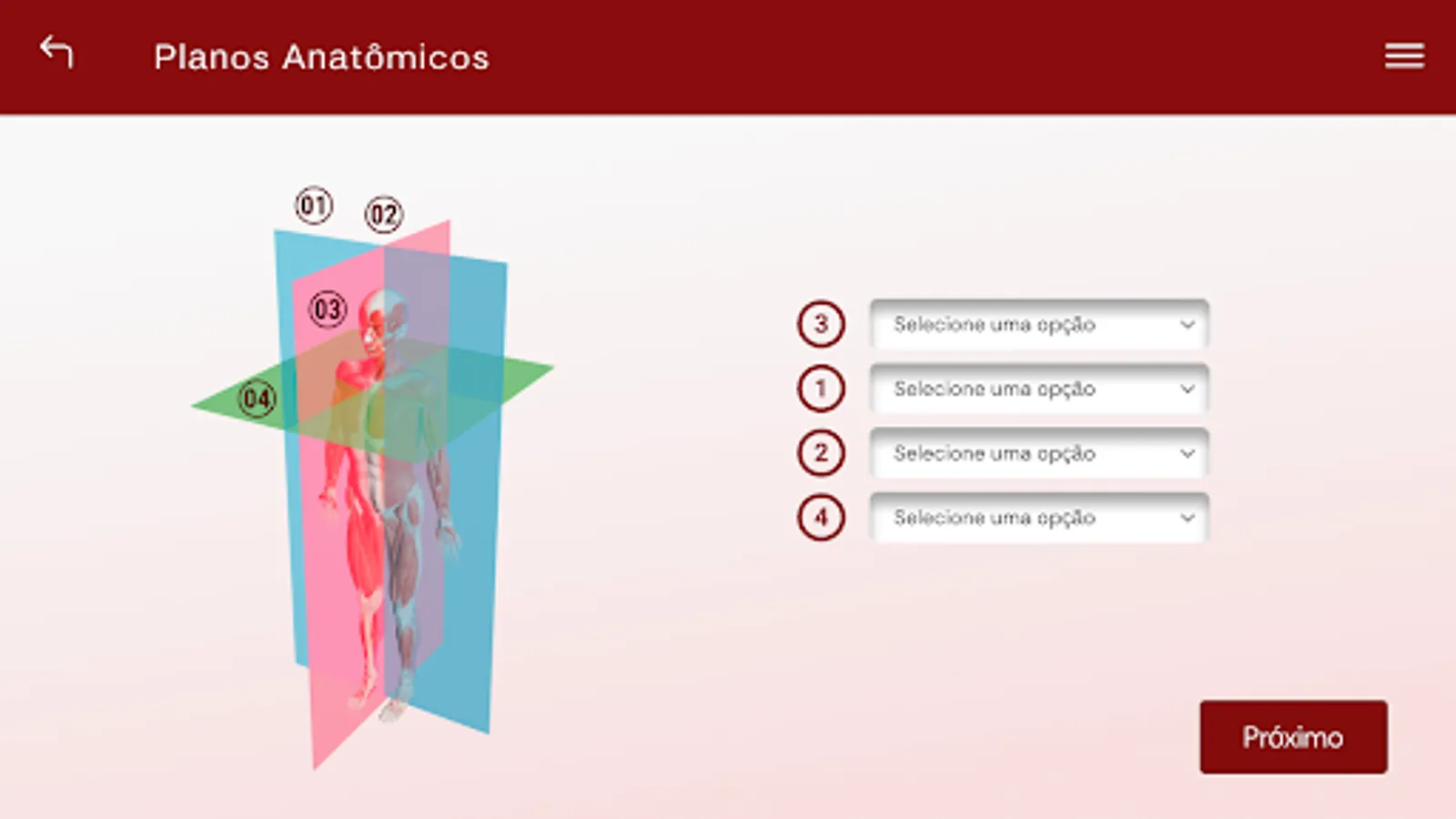
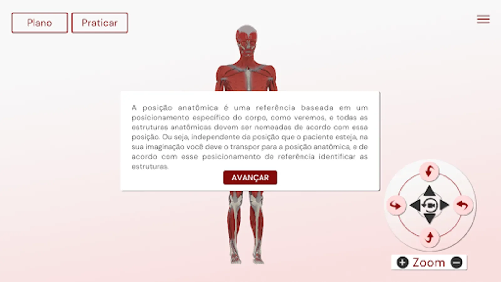
For a better understanding of the human body in the areas of anatomy and imaging diagnosis, it is important to know the anatomical planes and axes (sections). From this experiment, you will deepen your knowledge of important anatomical concepts.
At the end of this experiment, you should be able to:
Identify the purpose of using anatomical planes and axes in imaging exams;
Differentiate the main anatomical planes and axes in anatomical pieces;
Point out the anatomical axes present in a computed tomography.
2. WHERE TO USE THESE CONCEPTS?
Knowledge about the main planes and anatomical axes is fundamental for working in the area of diagnostic imaging, as it is from this information that the biomedical professional will properly position the patient in the equipment, as well as correctly capture the images requested by the team. doctor.
3. THE EXPERIMENT
This experiment will allow you to become familiar with anatomical structures that are constantly subjected to imaging examinations. From this, you will learn to identify the anatomical planes and axes of human organs, which will give you confidence in professional practice in the area of diagnostic imaging.
4. SECURITY
In the main diagnostic imaging centers, to carry out this experiment, it is essential that you use personal protective equipment (PPE) such as a lab coat, procedure gloves, long pants, closed shoes and tied hair up. In cases of units that work with ionizing radiation, it is also essential to use a dosimeter and a lead vest.
5. SCENARIO
The experiment will be carried out in the diagnostic imaging laboratory, where wet and/or synthetic anatomical parts of the brain will be available on a bench, in different planes and anatomical axes. In another part of the laboratory, computers will be available with internet access and the results of a patient's computed tomography exam, with the respective images, for you to evaluate.
In this laboratory, you will study important aspects of diagnostic imaging, taking into account fundamental concepts of anatomy and imaging, as biomedical professionals working in this area must have extensive knowledge of these sciences in their professional practice. The human body is made up of cells, tissues and organs that are grouped into organic systems. Although the body presents similarities between individuals of the same species, we can say that each individual is unique.
For a better understanding of the human body in the areas of anatomy and imaging diagnosis, it is important to know the anatomical planes and axes (sections). From this experiment, you will deepen your knowledge of important anatomical concepts.
At the end of this experiment, you should be able to:
Identify the purpose of using anatomical planes and axes in imaging exams;
Differentiate the main anatomical planes and axes in anatomical pieces;
Point out the anatomical axes present in a computed tomography.
2. WHERE TO USE THESE CONCEPTS?
Knowledge about the main planes and anatomical axes is fundamental for working in the area of diagnostic imaging, as it is from this information that the biomedical professional will properly position the patient in the equipment, as well as correctly capture the images requested by the team. doctor.
3. THE EXPERIMENT
This experiment will allow you to become familiar with anatomical structures that are constantly subjected to imaging examinations. From this, you will learn to identify the anatomical planes and axes of human organs, which will give you confidence in professional practice in the area of diagnostic imaging.
4. SECURITY
In the main diagnostic imaging centers, to carry out this experiment, it is essential that you use personal protective equipment (PPE) such as a lab coat, procedure gloves, long pants, closed shoes and tied hair up. In cases of units that work with ionizing radiation, it is also essential to use a dosimeter and a lead vest.
5. SCENARIO
The experiment will be carried out in the diagnostic imaging laboratory, where wet and/or synthetic anatomical parts of the brain will be available on a bench, in different planes and anatomical axes. In another part of the laboratory, computers will be available with internet access and the results of a patient's computed tomography exam, with the respective images, for you to evaluate.
AGLV1199 CORTES ANATÔMICOS Screenshots


Tap to Rate: